top of page
Imaging Techniques
To obtain highest quality and impact images for presentation and publication
Get the best images from your microscope

Bright Field
For standard stained slides

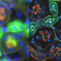
Fluorescence
To visualize fluorecently stained samples or those with fluorescing proteins and transgenic organisms

Stereo Fluorescence
fluorescence of larger samples, tissues, organs and whole animals

Confocal
To bring out the in focus focus of your sample while eliminating the out of focus components
_cells%2C_phas.jpg)
Phase Contrast
The simplest wayt see unstained portions of your sample, especially useful in cell culture applications

DIC and PlasDIC
Higher grade images of unstained samples can be obtained using DIC or PlasDIC techniques
Deconvolution and other Optical Sectioning Methods
Obtain excellent optical sections without the required investment in a confocal
bottom of page